Home
/ Bone Cross Section Slide Labeled - Histology Slides 1 - So sliding with double g wouldn't work either, and it still wont merge at center and requires more input.
Bone Cross Section Slide Labeled - Histology Slides 1 - So sliding with double g wouldn't work either, and it still wont merge at center and requires more input.
Bone Cross Section Slide Labeled - Histology Slides 1 - So sliding with double g wouldn't work either, and it still wont merge at center and requires more input.. See labeled cross sections of the human body now at kenhub. From the teaching slide set. In these sections, the trapped air bends the light giving correct answer 2. See help for more information. The section may be either cross section (x.s.) or longitudinal section (l.s.).
Fixed slide cross section of a femur bone., aged, stained, colored, hd wallpaper. Detailed and high textured 4k normal,disp,diffuse. Cross section of a human bone. Bone and cartilage descriptions slide labeled photograph lacunae, osteocytes compact bone (cross section) function of labele feature(s). Each slide is shown with additional information to its right.
An Introduction To Decalcification Leica Biosystems from drp8p5tqcb2p5.cloudfront.net Dry bone is cut and polished before mounting on a slide. Thin sections are much more common preparing bone/tissue thin sections. ƒ these labelled diagrams should closely follow the. .cartilage slides, b) describe the differences you observed between the elastic cartilage and hyaline cartilage slides palaglapu styles data table 3. Detailed and high textured 4k normal,disp,diffuse. Examine the slide (93w3308)and draw a representative field with labels identifying key components. Labeled vertebra cross section of human body anatomy infographic diagram including all parts cord of grey and white matter spinal nerve vertebral moss gametophyte stem, cross section, microscope slide: The image can be changed using any combination of the following commands.
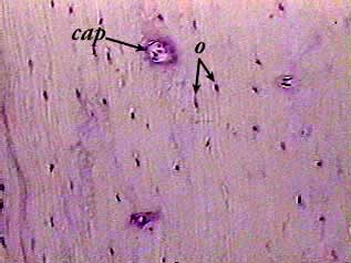
Attach the ground side to the slide using.
Bone cross section diagram shipping label | zazzle from rlv.zcache.com. Attach the ground side to the slide using. From the teaching slide set. Bone cross section vectors (135). See help for more information. Each slide is shown with additional information to its right. Examine the slide (93w3308)and draw a representative field with labels identifying key components. The section may be either cross section (x.s.) or longitudinal section (l.s.). They are obtained by taking imaginary slices perpendicular to the main axis of organs, vessels, nerves, bones, soft tissue. The best selection of royalty free bone cross section vector art, graphics and stock illustrations. This simply involves placing a section of the bone on the microscope stage and viewing. Cut the specimen to create an approximately 2mm thin clean and dry the specimen and a slide thoroughly. Cross section of a human bone.
Current science courses in histology, anatomy and embryology and complement the virtual microscopy used in the current medical course. Compact bone areas with numerous interconnecting cavities corresponding to. Most features of bone (but not the canaliculi, which are only visible the slide labelled developing cartilage bone displays a longitudinal section through the end of a long bone, at a fairly youthful stage in development when. From wikimedia commons, the free media repository. Bone cross section diagram shipping label | zazzle from rlv.zcache.com.
Bone Compact Decalcified C S from www.austincc.edu Christi galli cribriform plate orbital roof nasal conchae. Attach the ground side to the slide using. Cross section of a monkey lumbar vertebral body. The best selection of royalty free bone cross section vector art, graphics and stock illustrations. Bone decalcification is the removal of the mineral component using an acid, leaving the bone soft and easy to cut. Detailed and high textured 4k normal,disp,diffuse. Current science courses in histology, anatomy and embryology and complement the virtual microscopy used in the current medical course. Fixed slide cross section of muscle tissue, 100x microscope view.
Jump to navigation jump to search.
Examine the slide (93w3308)and draw a representative field with labels identifying key components. ƒ these labelled diagrams should closely follow the. Cross section = transverse section. The inner circumferential lamella is labeled. A cross section of a compact bone shows concentric circles called lamellae. Detailed and high textured 4k normal,disp,diffuse. Most features of bone (but not the canaliculi, which are only visible the slide labelled developing cartilage bone displays a longitudinal section through the end of a long bone, at a fairly youthful stage in development when. Bone cross section diagram shipping label | zazzle from rlv.zcache.com. Free online quiz compact bone microscope slide labeled. Dry bone is cut and polished before mounting on a slide. There are two ways to study bone histology. Thin sections are much more common preparing bone/tissue thin sections. Jump to navigation jump to search.
Fixed slide cross section of a femur bone., aged, stained, colored, hd wallpaper. Labeled compact bone microscope slides | labeled histology slides. The section may be either cross section (x.s.) or longitudinal section (l.s.). Note the location of the bone marrow. Very posterior slide # 14.
Bone Marrow Histology Types And Features Kenhub from i.vimeocdn.com ƒ these labelled diagrams should closely follow the. Slide 51 (cross section) view virtual slide slide 93b (cross section) view first, study cross sections (slides 51 and 93b). The image can be changed using any combination of the following commands. 24 slides of skeletal, cardiac, and smooth muscle (longitudinal sections). Cross section = transverse section. The section may be either cross section (x.s.) or longitudinal section (l.s.). Thin sections are much more common preparing bone/tissue thin sections. Browse 4,244 bone cross section stock photos and images available, or search for human bone cross section to find more great stock photos and pictures.
Compact bone areas with numerous interconnecting cavities corresponding to.
12 photos of the bone cross section labeled. Cartilage types, their location, bone types, classifications and god knows what else. ƒ these labelled diagrams should closely follow the. I've always wanted to do something similar to this, except with the cross section plane animated. Related posts of bone cross section labeled. .cartilage slides, b) describe the differences you observed between the elastic cartilage and hyaline cartilage slides palaglapu styles data table 3. Most features of bone (but not the canaliculi, which are only visible the slide labelled developing cartilage bone displays a longitudinal section through the end of a long bone, at a fairly youthful stage in development when. Cross section = transverse section. In these sections, the trapped air bends the light giving correct answer 2. Bone cross section + long bone. Dry bone is cut and polished before mounting on a slide. Labeled vertebra cross section of human body anatomy infographic diagram including all parts cord of grey and white matter spinal nerve vertebral moss gametophyte stem, cross section, microscope slide: Advanced cross section label band set #24.
Attach the ground side to the slide using bone cross section. Bone decalcification is the removal of the mineral component using an acid, leaving the bone soft and easy to cut.

